Research
Model-based Reconstruction for Dedicated Breast CT Perfusion Imaging
We aim to extract quantitative and functional information from high-resolution tomographic images of the breast by developing a dynamic contrast-enhanced breast CT modality. This has the potential to provide a large amount of functional information regarding breast tumors, well beyond only its morphology, which is key to characterize the biological profile of the tumor and its response during therapy. To reach the goal of quantitative breast CT imaging, image reconstruction plays an essential role, a task that we are addressing by developing a model-based iterative reconstruction algorithm. Even though reconstruction using direct inversion methods such as filtered backprojection (FBP) provides high quality images in a short time, it requires dense sampling in a specific geometry and extensive pre-corrections of the projection data in order to account for the assumptions that these data are continuously sampled, scatter-free, and mono-energetic. Iterative reconstruction, on the other hand, while slow compared to FBP, is much more flexible in reconstructing non-ideal data by allowing accurate modeling of the physical imaging process and the direct inclusion of external a priori knowledge in the reconstruction process. The methods developed to handle projection data with different physical properties can also be applied to allow for more flexible acquisition techniques that provide good quality images at lower dose levels. For example, a possibility can be alternating high and low energy acquisitions as used in dual energy imaging, and combining both in a mono-energetic image with a higher image quality compared to a single spectrum scan at the same dose level, effectively combining the high contrast from the low energy image and the low noise from the high energy scans.
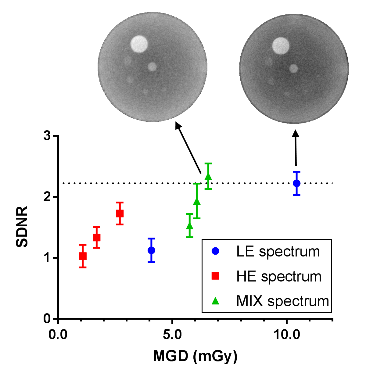
With our advanced reconstruction, we can reach the same image quality (SDNR - Signal Difference to Noise Ratio) as the current standard clinical acquisition with a low energy (LE) spectrum by mixing in projections with a high energy (HE) spectrum, at a lower total combined x-ray dose.
Researchers: