Research
Image Quality Evaluation Test for Clinical Performance Prediction in Digital Mammography
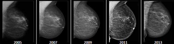
Image quality evaluation in mammographic screening is an important factor for optimizing early detection of breast cancer. Changes in image quality due to, for example changes in system design, could negatively affect the radiologist’s interpretation during mammogram reading. It is therefore important to understand what really affects the radiologist’s decision making and to have a method that can predict the clinical impact of changing any characteristic of the hardware or software of a system. Therefore, our group is developing a new VGA test able to predict clinical performance by basing its development and optimization on psychometric methods and its validation on ROC study results. This project, if successful, could lead to an objective evaluation of how suspicious lesions are interpreted by radiologists. For this project, we work together with radiologists and experts in this field from the Dutch Expert Centre for Screening (LRCB) and from other institutions across the Europe, such as the National Coordinating Centre for the Physics of Mammography (Guildford, UK), Cambridge University Hospital (Cambridge, UK), University of Lund (Mälmo, Sweden) and University of Leuven (Leuven, Belgium).
The impact on image perception of different post-processing mammographic algorithms. Which image is the best?
Researchers:
Joana Boita
Key Publications:
- J. Boita, A. Mackenzie and I. Sechopolous. "Validation of a method to simulate the acquisition of mammographic images with different techniques", 2019. Abstract. DOI.
- J. Boita, A. Mackenzie and I. Sechopoulos. "Breast phantom validation of a mammographic image modification method", 2018. Abstract. DOI.