Research
Image Denoising and Scatter Modelling and Correction
In x-ray imaging, several factors can negatively affect the image quality and potentially hide an abnormality, first among all the image noise produced by the random photon distribution within the image, and the x-rays scattering. Both effects can be limited by an accurate design and optimization of the imaging system, which can be achieved through model-based computer simulations, and through physical simulations with hardware phantoms. However, in most cases these effects cannot be completely avoided. The problem can therefore be addressed a posteriori, through the development of image processing algorithms able to correct noise and image non-uniformities after the image has been acquired. Within our group, we are developing image denoising and scatter correction algorithms mostly for tomographic breast imaging systems, such as dedicated breast CT and digital breast tomosynthesis. We work both with conventional image processing filtering, and with advanced patient-based scatter and noise simulations which are used to develop deep learning algorithms that can learn the correction process in a supervised fashion, for applications-specific correction.
With the development of dedicated image processing algorithm, scatter correction can be performed a posteriori, improving the quality of the image.
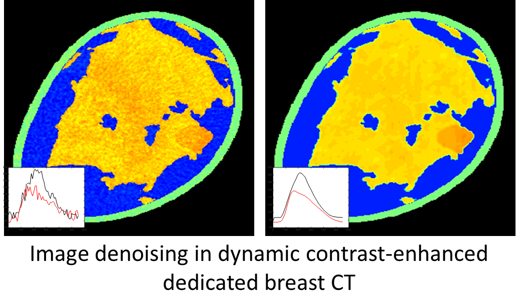
Example of results of a denoising filter based on Artificial Intelligence, able to recover both the quantum and the temporal noise.
Researchers:
Key Publications:
- K. Michielsen, C. Fedon, J. Nagy and I. Sechopoulos. "Dose reduction in breast CT by spectrum switching", 2018. Abstract. DOI.
- S. Ramamurthy, C. DÓrsi and I. Sechopoulos. "X-ray scatter correction method for dedicated breast computed tomography: improvements and initial patient testing.", 2016. Abstract. DOI.
-
Pautasso, J. J., Caballo, M., Mikerov, M., Boone, J. M., Michielsen, K., & Sechopoulos, I. (2023). Deep learning for x‐ray scatter correction in dedicated breast CT. Medical physics, 50(4), 2022-2036. Abstract. DOI.