Research
Image Analysis of High-Resolution CT Cochlea Images
A cochlear implant is a surgically implanted electronic device that provides a sense of sound to a person with severe to profound hearing loss. This device is implanted within the cochlea, the part of the inner ear involved in hearing. At the moment, a large variability still exists in speech understanding abilities after cochlear implantation. This could be related to the fact that cochlear implant electrodes are currently chosen independently from the patient anatomy of the inner ear, potentially resulting in a suboptimal positioning of the device. If accurate measurements of the cochlea could be performed pre-operatively, this could potentially allow to adapt the device to each single patient, and possibly improving the surgical outcome by reducing risk of intracochlear trauma and loss of residual hearing. The aim of this project (performed in cooperation with the Ear Nose Throat surgery department of Radboudumc) is therefore to develop and validate image analysis and deep learning algorithms to automatically perform complex measurements of the size of the cochlea from high-resolution CT images of patients.
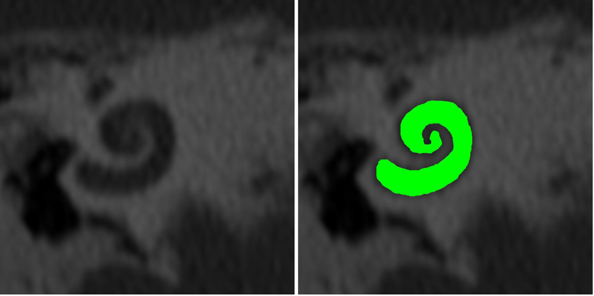
High-resolution CT cochlea image, and result of automatic segmentation.
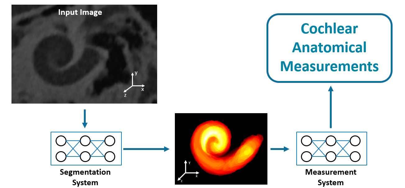
Scheme of the image analysis algorithm developed for automated cochlear segmentation and measurements.
Researchers:
Key Publications: